Congratulations! TingTing and Yaoping’s work on in-situ electroporation was reported in IEEE MEMS’ 2020 conference!
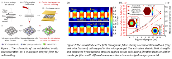
Labelling-assisted visualization is a powerful strategy to monitor the track of targeted cells for mechanism study, such as tumor metastasis. This work proposes an in-situ electroporation on an ultra-thin and high-porosity micropore-arrayed filter to introduce a fluorescent dye label into targeted cells. The whole procedure including cell separation and electroporation can be finished in a short duration (<15 min). High labelling efficiency (74.08±2.94%), along with a high viability (81.15±3.04% verified via live/dead staining) has been demonstrated with the rare (104) spiked mouse lung cancer cells after separated (efficiency at 81.47±2.17%) with a 10 μm micropore-arrayed filter from 1 mL whole blood. The achieved high viability is mainly attributed to the ultra-thin (10 μm) and high-porosity (46.79%), which contribute to decreasing forces (damages) applied on cells during filtration. This system demonstrated the applicability in all-in-one separation, labelling and retransfusion of targeted cells to monitor their track for tumor metastasis study.

